


Cultured mammalian cells / General staining
1. Mitochondria stained with lead citratein wet cells
Fully hydrated HeLa cells were fixed with 2% glutaraldehyde for 30min and stained with 1% Venables lead-citrate for 4 min. This image showsmitochondria (~300nm in width).
2. Wet Bovine sperm cells stained withosmium tetroxide
Fully hydrated Bovine sperm cells werefixed with 4% paraformaldehyde for 10 min and stained with 1% osmium tetroxide.
3. Wet CHO cells stained with uranylacetate
Fully hydrated CHO cells were fixed with 4%paraformaldehyde for 10 min and stained with 1% uranyl acetate.
Left: Lower magnification shows the overallstaining pattern with uranyl acetate.
Right: Structure of cell-cell contactsbetween neighboring cells at higher magnification.
4. Chromosomes stained with gold chloride
Cells grown in the QX-102 capsule were fixed with 2%Paraformaldehyde/1% Glutaraldehyde/PBS for 30 min. The cells were stained with0.1% gold chloride for 20 min and imaged in QX-102 Imaging Buffer. Left:NIH-3T3 cells, Right: CHO cells.
5. Macrophages
Macrophages (IC21) grown in QX-102 capsuleswere fixed and stained with uranyl acetate. Higher magnification image (right)shows ruffled borders of membranes. In collaboration with Prof. Paul Matsudaira(Head of MIT bio-imaging).
6. Cells stained with osmium tetroxide
Cells were grown in the QX-102 capsule, fixed (2%Paraformaldehyde/1% Glutaraldehyde/PBS) and stained with osmium tetroxide.
Left: NIH-3T3 cells
Right: Hela cells. Lipid droplets in thecytoplasm are seen as bright small spheres.
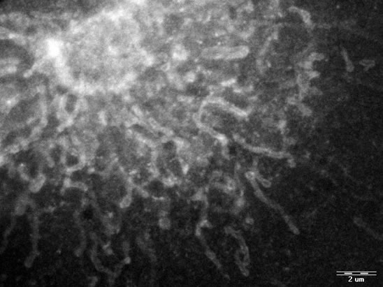
7. Mitochondria in HeLa Cells
Mitochondria visualization in HeLa cells that were fixed inGlutaraldehyde and stained with 2% Phosphotungstic acid. Mitochondria areeasily identified while their pleomorphic forms and structural variations areclearly seen.
9. Human blood cells
Left: Human blood plasma containingdifferent types of cells.
Middle: Polymorphonuclear human white bloodcells. After separation, cells were plated on the QX-102 capsule membrane.Efficient attachment and spreading of the population of interest were achieved.Fixation and consequent Uranyl Acetate and Osmium Tetroxide staining enhancedvisualization of cellular organelles, and highlighted the lipid bodies insidethe cytoplasm. These lipid bodies are known to be accumulated in circumstancessuch as inflammation.
Right: Red blood cells (Erythrocytes) wereplated on a poly-l-lysine coated QX-102 capsule membrane using a centrifuge at3000 rpm for five minutes. Cells were then fixed with 0.2% Glutaraldehyde andstained with 0.5% Osmium Tetroxide for 30 min.
10. Rat whole blood
Blood cells were attached to a poly-L-lysine coated QX-102 capsulemembrane and fixed with 0.2% Glutaraldehyde. Cells were stained with 0.5%osmium tetroxide for 30 min.